
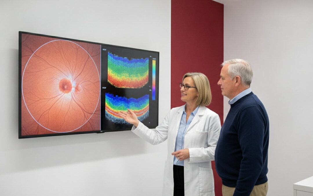
If you could see beneath the surface of your eye to spot a health issue years before it affects your vision, would you take that opportunity? When weighing up an oct scan vs retinal photography, it’s easy to feel overwhelmed by technical jargon. Most patients simply want to know they aren’t missing early signs of glaucoma or macular degeneration, yet they often feel uncertain about which technology offers the best protection or whether the NHS covers these advanced checks.
We believe that looking after your sight should be a warm, reassuring experience rather than a confusing one. This guide explains the critical differences between a surface map and a deep-tissue cross-section so you can understand exactly what each machine “sees.” You’ll gain the confidence to choose the right level of care for your eyes, ensuring you leave your next appointment at our Rayleigh or Benfleet centres with total peace of mind. We’ll break down the benefits of both imaging types, helping you protect your vision for the years to come.
Key Takeaways
- Learn how modern eye imaging goes beyond standard sight tests to identify health issues hidden beneath the retinal surface.
- Understand the critical difference between an oct scan vs retinal photography, including how one acts as a surface map whilst the other provides a 3D cross-section.
- Discover why OCT technology is vital for the early detection of conditions such as glaucoma and macular degeneration, often years before symptoms appear.
- Find out how the independent approach at our Rayleigh and Benfleet branches ensures you receive a thorough, unhurried consultation tailored to your eye health.
- Get clear guidance on the NHS status of these advanced scans so you can make an informed choice for your long-term vision and peace of mind.
Understanding Modern Eye Imaging in South Essex
For many years, the standard eye examination focused almost entirely on how well you could read a Snellen chart. If you could see the bottom line, your eyes were considered healthy. Times have changed. Modern optometry now prioritises what’s happening behind the pupil, using digital diagnostics to spot life-altering conditions before they impact your daily life.
At the heart of this diagnostic revolution are two distinct technologies. Digital Retinal Photography acts as a high-resolution camera for the back of your eye, whilst Optical Coherence Tomography (OCT) provides a detailed, 3D cross-section. Understanding the difference between an oct scan vs retinal photography is key to choosing the right level of care for your vision. By bringing this hospital-grade technology to our independent practices, we’re able to offer a level of detail once reserved for specialist clinics.
The Shift from Sight Testing to Eye Health Screening
Vision is only one piece of the puzzle. You can have perfect 20/20 sight and still have the early stages of a condition like glaucoma or macular degeneration. These “silent” conditions are often asymptomatic, meaning they don’t cause pain or noticeable vision loss until significant damage has already occurred. Early warning signs simply don’t exist for many of these issues, making the “wait and see” approach risky for long-term eye health. Preventative screening allows us to see the unseen. By monitoring the internal structures of the eye, we can identify microscopic changes in the retinal layers, ensuring that any intervention happens as early as possible.
Why Rayleigh and Benfleet Residents Choose Advanced Imaging
As an independent group established in the South Essex community since 1989, we’ve seen first-hand how technology changes lives. Our patients in Rayleigh and Benfleet value personal attention, and advanced imaging is a huge part of that. Having these scans on your local clinical record allows us to compare your eye health year-on-year with incredible precision. It’s about building a story of your eyes over time rather than just taking a snapshot.
This longitudinal data is invaluable. It helps us distinguish between a stable, lifelong quirk in your eye’s anatomy and a genuine cause for concern. By identifying these nuances in-house, we often reduce the need for stressful and unnecessary hospital referrals. At Castle Opticians, our commitment to investing in the latest diagnostic tools ensures that our neighbours receive the most thorough health check available on the high street, delivered with the warm hospitality of a trusted local expert.
Digital Retinal Photography: Capturing the Surface of Your Eye
Digital retinal photography uses a specialised device called a fundus camera to take a high-resolution photograph of the back of your eye. When comparing an oct scan vs retinal photography, it helps to think of this method as a “Surface Map.” It captures the layout of your retina, including the optic nerve, the macula, and the intricate network of blood vessels. When your optician reviews this image, they’re looking for subtle markers of health or disease. This might include retinal “freckles” known as nevi, tiny haemorrhages that could indicate blood pressure issues, or pigment changes that suggest early wear and tear.
The process is entirely non-invasive and takes only a few seconds. You’ll see a bright flash, similar to a standard camera, and the image appears on screen almost instantly. It’s a quick, comfortable part of a modern check-up that provides a wealth of information about your general wellbeing without any physical contact with the eye itself.
The Benefits of a Permanent Digital Record
One of the greatest advantages of this technology is the ability to create a historical record of your eye health. By storing these images digitally, we can perform side-by-side comparisons during your future visits. Spotting a minute change in a blood vessel or a pigment spot is much easier when we have the previous year’s “map” for reference. This level of detail is a cornerstone of a comprehensive eye test in Essex, where we prioritise long-term monitoring over quick snapshots.
These images are also incredibly useful for collaborative care. If we notice something that requires a second opinion, we can securely share your digital files with your GP or a local hospital specialist. This ensures everyone involved in your healthcare has the same clear view of your eyes. When Choosing the Right Method for screening, the ability to track changes over time is often the most compelling reason to include photography in your routine.
What Retinal Photography Can (and Cannot) Detect
Whilst photography is excellent for spotting surface-level anomalies and diabetic changes, it does have its limitations. Because it’s a two-dimensional image, it only shows the top layer of the retina. It’s very effective at showing us where a problem is on the surface, but it cannot tell us how deep that problem goes.
This is the primary distinction when comparing an oct scan vs retinal photography. A “clear” photo is a great sign, but it doesn’t always guarantee a 100% healthy eye. Some conditions, like early-stage wet macular degeneration, start as fluid leaks deep within the retinal layers, which might remain invisible on a standard photograph. If you’re concerned about specific symptoms or have a family history of eye disease, you might find that booking a thorough eye test with advanced imaging provides the extra layer of certainty you need.
OCT Scanning: Seeing Beneath the Retinal Surface
If retinal photography is the surface map, then OCT is the architectural blueprint of your eye. Optical Coherence Tomography uses near-infrared light waves to penetrate the tissue, acting much like sonar to map the internal structures. It’s often described as an MRI or ultrasound for the retina because it provides a high-definition, 3D cross-section. This technology allows our clinicians to peer through the surface and examine ten distinct layers of the retina individually. Identifying fluid, swelling, or thinning in these deep layers is where the choice between an oct scan vs retinal photography becomes vital for your long-term health. A clinical study comparing OCT and retinal photography confirms that OCT is far more sensitive at detecting subtle abnormalities that a standard 2D image simply cannot capture. By looking at the foundations of the eye, we can see health issues that are literally buried beneath the surface.
Early Detection of Glaucoma and Macular Degeneration
Early intervention is the single most important factor in preserving your sight as you age. With glaucoma, damage often begins at the microscopic level within the retinal nerve fibre layer (RNFL). By measuring the thickness of these nerves in microns, we can spot the very first signs of thinning before you notice any change in your vision. Similarly, OCT is the gold standard for managing Age-Related Macular Degeneration (AMD). It allows us to distinguish between “dry” AMD and the more urgent “wet” AMD with incredible precision. Seeing fluid build-up beneath the surface means we can act immediately, often years before a standard sight test would raise an alarm. When comparing an oct scan vs retinal photography, the ability to see beneath the surface is the clear winner for early diagnosis. This proactive approach is why we recommend enhanced screening for all our patients over forty.
The Power of Baseline Scans and Trend Analysis
When you visit our Rayleigh or Benfleet practices, we use OCT to create a “biological fingerprint” of your eye structure. Our software doesn’t just take a picture; it performs a complex trend analysis over time. It tracks changes at a micron level across your visits, comparing your current scan to every previous one on your record. This allows us to see if a particular area is thinning or if a tiny pocket of fluid is starting to form. OCT is a micron-resolution imaging technology that identifies pathology before it impacts vision. This level of detail gives you and your family the ultimate peace of mind, knowing that your eye health is being monitored by a trusted local expert with the best tools available.
OCT Scan vs Retinal Photography: Which Do You Need?
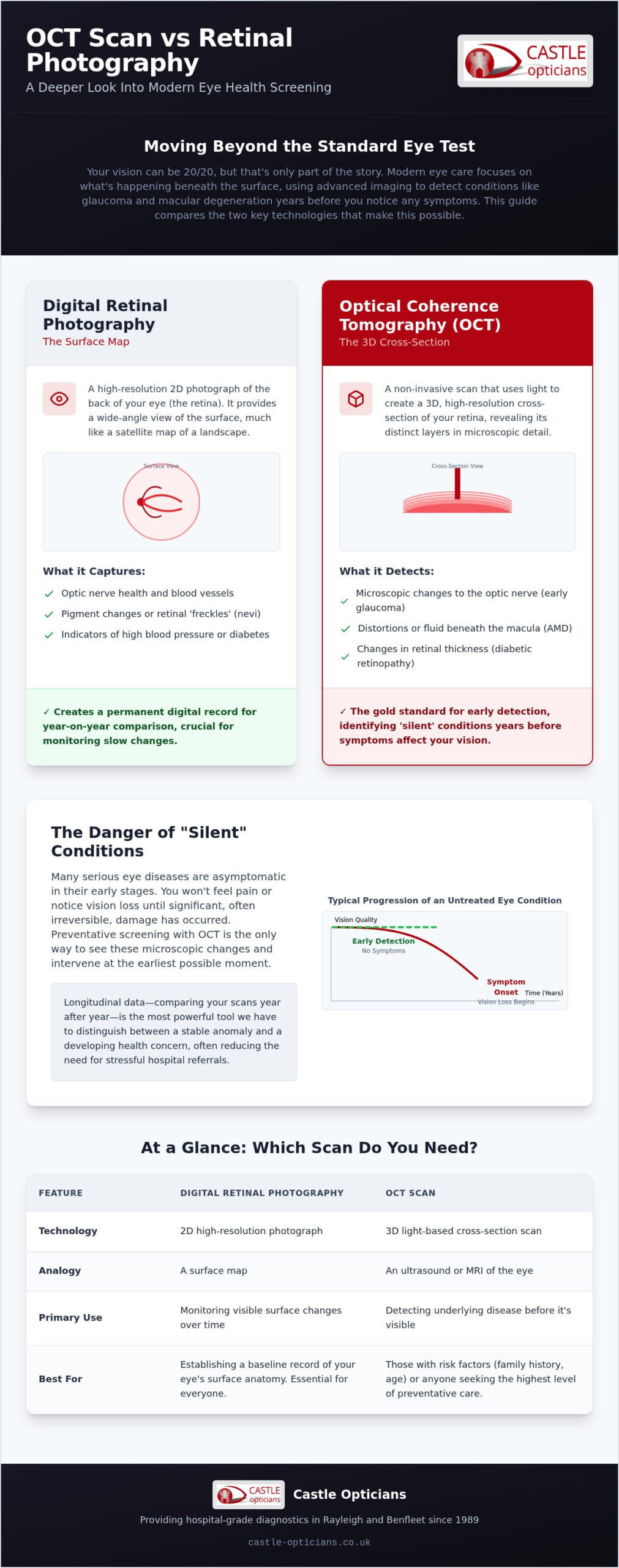
Choosing between an oct scan vs retinal photography is best understood through a simple analogy. Retinal photography is like a high-quality map of a house; it shows you the layout of the rooms and where the furniture is placed. An OCT scan is more like a full building inspection. It looks behind the wallpaper and beneath the floorboards to check the structural integrity and foundations. Whilst the map tells you where you are, the inspection tells you if the building is truly sound. Both have a specific role in your eye care, but they offer very different levels of insight.
| Feature | Retinal Photography | OCT Scan |
|---|---|---|
| Depth of View | 2D Surface Image | 3D Cross-Sectional layers |
| Primary Use | Mapping surface changes | Detecting structural disease |
| Disease Focus | Diabetes, freckles, pigment | Glaucoma, AMD, internal fluid |
| Recommended For | All routine patients | Over 40s, family history, symptoms |
For a young, healthy patient with no family history of eye disease, a standard retinal photograph may be sufficient to document their current eye health. However, we strongly prioritse OCT for anyone over the age of forty, those with high spectacle prescriptions, or individuals with a family history of glaucoma. These groups are at a higher risk of structural changes that occur deep within the tissue, where a 2D photograph simply cannot reach.
A Complementary Approach: Why Castle Opticians Uses Both
We don’t view this as a choice of one or the other. In fact, these technologies work best when used together. The retinal photograph provides the “big picture” context, allowing us to see the overall health of the blood vessels and optic nerve head. The OCT then allows us to “zoom in” on specific areas of concern to see what is happening beneath that surface. This dual-layered approach is the foundation of a comprehensive eye health check in Essex. By combining both, we tailor our diagnostic approach to your specific history, ensuring no detail is overlooked.
Addressing the Cost vs. Value Misconception
In the UK, a standard NHS eye test is designed to check your basic vision and obvious signs of disease. Because OCT technology is incredibly advanced, it’s typically offered as a private supplement to your NHS or private examination. Whilst there’s a small additional fee, the value lies in the long-term protection of your sight. Identifying a condition like glaucoma years earlier can mean the difference between simple daily eye drops and significant vision loss. We believe in helping our patients make an informed choice for their health, providing clear guidance without any hard-sell pressure. If you’re ready to see your eye health in more detail, you can book your next consultation at our Rayleigh or Benfleet branches today.
Advanced Diagnostics at Castle Opticians: Rayleigh and Benfleet
Since 1989, Castle Opticians has been a fixture of the South Essex community, providing a level of personal care that only an independent practice can offer. We believe that technology is most effective when paired with time and unhurried expertise. When you visit our Rayleigh or South Benfleet branches, you aren’t just another booking in a crowded diary. You’re a neighbour who deserves a thorough understanding of their vision. Our clinical team takes pride in explaining the complex data from your oct scan vs retinal photography in plain British English, ensuring you leave your appointment feeling informed and looked after rather than confused by technical jargon.
An advanced imaging appointment at our centres is designed to be a calm, hospitable experience. We’ve invested in hospital-grade equipment to ensure our patients have access to the best possible diagnostics right on their local high street. This commitment to excellence has defined our service for over three decades, and it continues to guide how we look after the generations of families who trust us with their sight.
The Independent Advantage: Time to Care
One of the most significant benefits of choosing an independent optician is the gift of time. Unlike corporate high-street chains that often operate on restrictive, high-volume schedules, we offer longer consultations. This allows our optometrists to perform a deeper analysis of your scans and answer any questions you might have about your long-term eye health. It also means you’ll often see the same friendly face for your follow-up appointments, providing a continuity of care that is vital for tracking subtle changes in your retinal structure.
Our expertise doesn’t stop at clinical diagnostics. Once we’ve ensured your eyes are healthy, our team is on hand to help you find the perfect vision correction. We offer a curated selection of spectacle frames in Rayleigh and South Benfleet, combining local style with high-quality lenses tailored to your lifestyle. It’s a complete, start-to-finish service delivered with a personal touch.
Next Steps for Your Eye Health
Preparing for your advanced imaging is entirely stress-free. There are no needles, no pain, and the imaging process itself doesn’t involve the uncomfortable “puffs of air” often associated with older eye tests. You simply lean forward, look at a target light, and let the technology do the rest in a matter of seconds. We believe in transparency, which is why we discuss your results with you immediately. There’s no waiting for a letter in the post; you’ll see your retinal maps and 3D cross-sections on screen whilst your optician guides you through what they mean.
Your vision is precious, and early detection is the best way to protect it for the future. Whether you have a specific concern or simply want the peace of mind that comes with a professional health check, we’re here to help. Book your advanced eye examination at Castle Opticians today and experience the difference that independent, expert care makes to your wellbeing.
Protecting Your Vision for the Future
Understanding the critical differences between an oct scan vs retinal photography is the first step toward truly proactive eye health. Whilst digital photography provides a vital surface map of your retina, an OCT scan offers the architectural depth required to spot conditions like glaucoma or macular degeneration years before symptoms appear. Since 1989, our independent, family-run practice has prioritised this level of clinical detail for our neighbours across Rayleigh, Benfleet, and Canvey Island. We believe that everyone deserves access to hospital-grade technology without the sterile atmosphere of a large clinic.
By choosing a local expert, you benefit from longer consultations and a team that genuinely cares about your long-term wellbeing. We take the time to explain your results clearly, ensuring you leave with total confidence in your care. Your sight is one of your most precious assets, and we’re here to help you safeguard it with the most advanced tools available on the high street. Book your comprehensive eye test at Castle Opticians today and enjoy the peace of mind that comes with a thorough, professional health check. We look forward to welcoming you soon.
Frequently Asked Questions
Is an OCT scan painful or uncomfortable?
An OCT scan is completely painless and non-invasive. You simply sit in front of the machine and look at a target light whilst the scanner captures images using light waves. There are no needles, no physical contact with the eye, and unlike some older tests, there are no uncomfortable puffs of air. It’s a quick and relaxing process that feels very similar to having a standard photograph taken.
Does the NHS cover the cost of an OCT scan?
Standard NHS sight tests do not typically cover the cost of an OCT scan as they are considered an advanced diagnostic supplement. Whilst your basic eye test may be funded by the NHS, the enhanced imaging is usually offered as a private add-on service. This allows us to use hospital-grade technology to provide a more thorough health check than the standard framework requires, giving you extra peace of mind.
How often should I have an OCT scan vs retinal photography?
Most patients should have these scans every one to two years as part of their routine eye examination. If you are over forty or have a family history of conditions like glaucoma, we may recommend more frequent checks to monitor for subtle changes. Regular imaging is essential for building a historical record, allowing our experts to perform a detailed comparison of your oct scan vs retinal photography results over time.
Can an OCT scan detect things that a standard eye test misses?
Yes, an OCT scan can identify structural changes in the deep retinal layers that a standard eye test might miss. By providing a 3D cross-section, it reveals issues like microscopic fluid leaks or thinning of the nerve fibre layer long before they become visible on the surface. This early detection is the primary reason we recommend advanced imaging for anyone concerned about “silent” conditions that develop without symptoms.
Do I need to have my pupils dilated for these scans?
Pupil dilation is not usually required for these advanced scans. Our modern equipment is designed to capture high-resolution images through most natural pupil sizes. However, if your pupils are particularly small or if you have certain types of cataracts, your optician might suggest using dilating drops to ensure the clearest possible view. If this is necessary, we will always discuss it with you during your consultation at our Rayleigh or Benfleet branches.
What happens if the OCT scan finds something abnormal?
If an abnormality is found, your optician will discuss the results with you immediately in plain English. Depending on the findings, we might suggest more frequent monitoring to track the condition or, if necessary, arrange a referral to a hospital specialist for further treatment. Because we are an independent practice, we take the time to ensure you understand the next steps and feel supported throughout the entire process.
Is retinal photography still necessary if I have an OCT scan?
Retinal photography remains a vital tool even if you choose an OCT scan. Photography provides a wide-field “surface map” of the eye, which is excellent for documenting the appearance of blood vessels and pigment. When comparing an oct scan vs retinal photography, it’s best to view them as complementary technologies. Using both ensures we have both a clear overview of the surface and a deep architectural view of the eye’s foundations.
How long does the advanced imaging process take during an eye test?
The advanced imaging process is very efficient and usually takes less than five minutes to complete. Capturing both the retinal photographs and the 3D OCT scans is a seamless part of your enhanced eye test. This small investment of time allows our clinical team to gather a vast amount of data about your eye health, which we then review and explain to you during the remainder of your unhurried consultation.